Constraints
Constraints derived from over 250 experimental findings correlated with behaviors indicative of the generation of first-person properties are examined. Of these, nearly 50 were used to systematically restrict the space of a unique viable solution capable of generating both behavior and first-person properties. It is expected that constraints from the remaining findings will reveal features enabling seamless interconnections.
How hidden is the solution & how do the constraints help to find it?
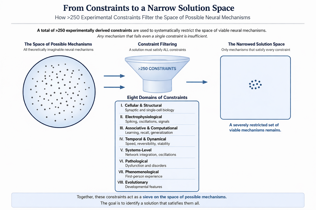
The disparate findings from eight domains of the system functions (given after the next figure) are expected to be interconnected through a novel mechanism (Fig.2), analogous to an adaptor protein that binds to multiple molecular partners. This explains how the unique properties of the solution enable large number of features of the system observed from different levels to get integrated in an interconnected manner.
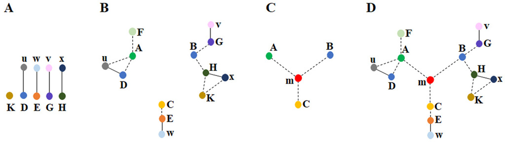
Figure 2. Disparate findings of the system in different levels are interconnected with each other through a unique solution A) The features of the system are detected either directly (represented by the capital letter K) through our sensory systems or indirectly (represented by the capital letters D, E, G, H) through findings such as protein staining or behavioral observations. These features are connected through smaller letters (u, w, v, x), which represent relationships between the features (e.g., the observation of u enables the detection of D). B) By utilizing both commonly employed direct and indirect methods, three clusters of interconnected findings (depicted by dotted lines) are identified across different levels (observations from various fields of brain science). In many instances, it is not possible to establish connections between these clusters. For example, no direct connection was found between: 1) the changes in learning and the internal sensations of memory occurring at millisecond timescales, and 2) sleep & long-term potentiation (LTP). However, by examining the constraints within each cluster, we can assess whether they can be unified through a common operational mechanism. In the context of the nervous system, a vast array of findings and the constraints they provide can be explored. C) By leveraging the constraints from certain features within each cluster of seemingly unrelated findings (e.g. A, B, C), it may become possible to derive a deep underlying principle (a structure-function solution m) that enables their interconnection. This solution is expected to offer a mechanism for generating internal sensations at millisecond timescales. D) The solution m provides an explanation of how various findings within each cluster are interrelated with one another & with findings from other clusters, as shown in B). The ability of solution m to integrate findings across all clusters makes it a further verifiable solution (Fig. from Vadakkan KI (2019) Phys Life Rev.31:44-78.
| Domain 1 Cellular and Structural | |
Finding | Constraint (The solution must be able to provide interconnected explanations for all the constraints) |
All the stimuli during learning & memory retrieval propagate through synaptically-connected neuronal circuits. | Mechanism should operate synchronously with the synaptically connected circuitry. |
Any subset of 140 input signals reaching the axonal hillock is sufficient to fire a neuron (Palmer et al., 2014; Eyal et al., 2018) | This extreme degeneracy suggests that the information storage mechanism should be taking place at the level of the input terminals. |
Spine neck resistance (Tønnesen et al., 2014) and attenuation of potentials as they propagate along the dendrite towards the soma (Spruston, 2008). | The ideal region for learning changes to take place is the spine head region. |
Optogenetic activation of presynaptic inputs in lateral amygdala (LA) forms associative fear memory (Kwon et al., 2014). | LA is a region where inputs from two associated stimuli converge. Hence there should be convergence of inputs at the synaptic level. |
Fear learning generates local connectivity between lateral amygdala (LA) neurons (Abatis et al., 2024) as evidenced by depolarization in a small subset of neighboring LA neurons, when a single LA neuron is stimulated. | During cued fear conditioning, lateral connections are expected to occur between LA neurons. These are expected to occur between the dendritic spines of these neurons. |
Since associatively learned inputs synapse on to the spines of LA neurons, the inter-neuronal interaction is expected to take place between the synapses. | Since the minimum statement to say that a synapse is activated is generation of postsynaptic potentials, minimum interaction by which this can be accomplished is through inter-spine interaction. |
Synapse-dense cortical areas with tightly packed neuropil have adjacent spines (Kasthuri et al., 2015; Zhu et al., 2021; Gemin et al., 2021). | This increases the probability for inter-neuronal inter-spine interactions. |
The dendritic arbors of pyramidal neurons display significant territorial overlap (Mizuseki et al., 2011; Bezaire & Soltesz, 2019; Iascone et al., 2020). | This increases the probability of inter-neuronal inter-spine interactions. |
The sister branches on a neuron’s dendritic tree often avoid overlapping (Grueber & Sagasti, 2010). | This increases the probability of inter-neuronal inter-spine interactions. |
The mean inter-spine distance on the dendrite of a pyramidal neuron exceeds the mean spine diameter (Konur et al., 2003). | This allows interaction between the spines that belong to different neurons. |
Over half of the spine surface area lacks ensheathment by astrocytic processes (Ventura & Harris, 1999). | The remaining half of the spine surface area is free to interact with the abutted spine surfaces of other neurons. |
Synapses devoid of astrocytic coverage emerge in the amygdala during the consolidation of Pavlovian threat conditioning (Ostroff et al., 2010). | The reduction or disappearance of astrocytic pedicels during the consolidation may increase the probability of inter-neuronal inter-spine interaction. |
Most excitatory glutamatergic synapses are located on dendritic spines, which enlarge during learning. Glutamate induces spine enlargement in both hippocampal slices (95%) (Matsuzaki et al., 2004) & the neocortex in vivo (22%) (Noguchi et al.,2019). | Spine enlargement can both increase the surface area and also displace the extracellular matrix (ECM) increasing the probability for inter-neuronal inter-spine interaction. |
Fear conditioning is associated with enlarged synapses on the dendritic spines of LA neurons (Ostroff et al., 2010; Choi et al., 2021). | Synapse enlargement can result from the expansion of either pre- &/or postsynaptic terminals. Since inter-spine interaction is an anticipated learning change, this matches. |
Motivation enhances learning & is associated with the release of dopamine, which activates dopamine receptors in various regions of the brain (lino et al., 2020). | Dopamine is known cause spine expansion (Yagishita et al., 2014). Abutted spines from different neurons, when expanded can undergo certain inter-spine interactions. |
Contextual fear conditioning recruits newly synthesized GluA1-containing AMPA receptors into the spines of hippocampal memory-ensemble cells in a learning-specific manner (Matsuo et al., 2008). | Exocytosis of vesicles adds vesicle membranes segments to the spine membrane increasing spine size, which can facilitate inter-spine interactions. |
Autophagy leads to memory destabilization and the erasure of auditory fear memories, a process associated with AMPA receptor endocytosis (Shehata et al., 2018). | Endocytosis of vesicles removes vesicle membranes segments from the spine membrane decreasing spine size, which can separate interacting spines of different neurons. |
An inhibitor of AMPA receptor endocytosis partially rescued long-term memory deficits in mice with elevated levels of amyloid-β (Yan et al., 2024). | Preventing endocytosis of vesicles prevents removal of vesicle membranes segments from the spine membrane & prevent decrease in spine size, maintaining abilities for inter-spine interactions. |
Mice injected with histone acetyltransferase (HAT) exhibit enhanced fear memory. Neurons in which HAT was overexpressed are part of the engram (Santoni et al., 2024). Removal of histone protein from DNA enhanced fear memory. The study found that a) neurons in which HAT is over-expressed are the neurons that fire during memory retrieval, & b) optogenetic silencing of these specific set of neurons prevents fear memory recall. | Removal of histone proteins from DNA facilitates the gene expression, whose protein products can be used for the inter-neuronal inter-spine interaction mechanism. HAT over-expressed neurons allow more inter-spine interactions to form during learning. These excess inter-spine interactions help to generate robust first-person property of fear and behavioral motor actions reminiscent of fear. |
Most learning events result in a working memory that lasts only for a short period. All long-term memories has had working memories immediately after learning. | Inter-spine interaction is a highly reversible process. Inter-spine interactions extending to inter-membrane hemifusion events can stabilize the inter-spine interactions variable periods. |
The capacity to store extensive sets of learning-induced changes underlies the ability to retrieve a large number of behavioral expressions during memory retrieval events. Using a finite number of dendritic spines this must be achieved. | A single spine that can interlink with more than one spine can lead to the formation of islets of interlinked spines (IILSPs). This can allow a specific cue stimulus can reactivate specific sets of interlinked spines to generate corresponding sets of behaviors. |
Higher brain functions occur only when the frequency of oscillating extracellular potentials falls within a narrow range, as evidenced by EEG recordings (Rusalova, 2006). | Oscillations needs vector components that contribute to it. Since all cortical neurons have their apical dendritic terminal attached to the inner pial surface, large number of inter-spine interactions can be formed. Synaptic transmission in mostly vertically oriented synapses & horizontally oriented inter-spine interactions can contribute vector components for oscillations. |
Even though extracellular recordings like EEG is biased by filtering effects (e.g. extracellular tissue act as a low-pass filter), brain operates in a narrow range of frequencies of EEG waveforms recorded from specific locations. For e.g. alpha rhythm (8–12 Hz) recorded from occipital cortex during relaxed wakefulness (Niedermeyer & da Silva, 2017). | The operational mechanism is expected to generate vector components of the oscillating extracellular potentials, accompanied by corresponding intracellular ionic concentration changes within the neuronal processes of the involved neurons. Inter-spine interaction mechanism provides vector components for oscillating potentials. |
A disconnect between dendritic depolarization & neuronal firing has been observed during fear conditioning (d’Aquin et al., 2022). | An operational mechanism, most likely related to the first-person property, is expected to emerge at the dendritic level, independent of neuronal firing. |
| First-person arguments | |
| Memories are virtual first-person inner sensations. | It is essential to identify both the location & the mechanism responsible for its generation. |
| Memory retrieval occurs on a millisecond timescale. | Since the memory of a learned association can be retrieved immediately after learning, it is essential to demonstrate a retrieval mechanism that operates within milliseconds |
| After associative learning between two items, the presentation of one item triggers the memory of the other. | The learning mechanism must be capable of explaining how either item in an associative pair can serve as a cue to evoke the memory of the other. Therefore, the mechanism must inherently support bidirectional activation. |
| Loss of consciousness occurs during a generalized seizure & typically resolves once the seizure ends. | The mechanism of seizure generation should account for how the inner sensation of consciousness is lost. |
| Changes in consciousness are proportional to variations in the frequency of oscillating extracellular potentials that deviate beyond a narrow physiological range. | An explanation is needed regarding how the narrow range of oscillating extracellular potential frequencies is linked to the maintenance of normal consciousness. |
| Gradual changes in the qualia of the inner sensation of memory occur as the cue stimulus changes progressively. | It is expected that there is a mechanism to integrate individual elements in order to generate the inner sensation of memory. |
| Absence of cellular changes during memory retrieval. | Memory retrieval should involve the passive reactivation of changes that occurred during learning, which induces units of internal sensations. |
| Instant access to very large memory stores. | It is essential to explain how a vast number of memories can be retrieved instantly. This requires demonstrating an instantaneous combinatorial mechanism of unitary operations that occurs without delay. |
The inner experience of certain higher brain functions can occur without any accompanying motor actions. | The mechanism that generates inner sensations must be capable of demonstrating either that no behavioral motor actions are produced alongside a particular inner sensation, or that the motor action can be voluntarily suppressed. Motor should be able to get inhibited without inner sensations getting inhibited. |
| Domain 2 Electrophysiological a) Long-term potentiation | |
| Strength of LTP induced is correlated with the ability to learn (Matynia et al., 2002; Lynch MA, 2004). | Both learning and LTP are using comparable mechanisms. |
| Dopamine enhances both motivation-driven learning (Bromberg-Martin et al., 2010) & LTP (Otmakhova & Lisman, 1996). | Dopamine is known to cause increase in the size of spines (Yagishita et al., 2014). Inter-spine interaction can be influenced by the expansion of abutted spines. |
| An increase in the amplitude of miniature EPSPs (mEPSPs) occurs following LTP induction (Manabe et al., 1992). Amplitude of mEPSPs is thought to be influenced by an increase in the number or functional efficacy of AMPA receptors (Malenka & Nicoll, 1999). | The recording electrode is electrically connected to abutted spines through inter-spine interactions, allowing current from interlinked spines – primarily originating from different neurons – to contribute to the recorded signal. Current arriving from interlinked spines through the inter-spine interactions can explain the observed increase in mEPSP amplitude (Vadakkan, 2019). |
| Learning can be occluded following LTP induction, & conversely, LTP can be occluded after learning (Moser et al., 1998; Whitlock et al., 2006). | It is necessary to explain a mechanism that can take place both during learning and LTP induction. |
| Fusion blockers block LTP | These blockers must be capable of blocking intermediate stages of fusion between all the membranes. |
| LTP stimulation is associated with lateral movement of vesicles containing AMPA receptor subunits (Rumpel et al., 2005; Makino & Malinow, 2009; (Granger et al., 2013) & distribution of these receptors to the membranes (Shi et al., 1999; Passafaro et al., 2001). Blockade of exocytosis of these vesicles cause significant reduction in LTP (Kennedy et al., 2010; Ahmad et al., 2012). Endocytosis of AMPA receptors leads to LTP decay and memory loss (Dong et al., 2015). | Addition of large amount of vesicle membranes and AMPA receptor subunits to the spine membrane should be able to provide mechanistic explanations how they contribute to the induction of LTP after nearly 20 to 30 seconds. Similarly, it is necessary to show how this learning or LTP induced changes can be reversed by endocytosis of vesicles containing AMPA receptors. |
LTP is associated with the enlargement of dendritic spine heads (Lang et al., 2004). | Spine enlargement increases spine surface area and reduces the inter-spine extracellular matrix. This facilitates inter-neuronal inter-spine interactions. |
| Small spines have been identified as preferential sites for the cellular changes associated with LTP induction (Matsuzaki et al., 2004) . | LTP stimulation allows spines to expand, which in turn increases the probability for inter-neuronal inter-spine interactions necessary for the formation of large number of inter-spine interactions that allow connections from the stimulating to recording electrode when LTP is measured in response to a regular stimulus after LTP stimulation. |
| High-energy stimulation alone can bypass the requirement of lateral movement of vesicles containing AMPA receptor subunits (Makino & Malinow, 2009) for LTP induction. Instead, LTP can be directly induced even in the absence of AMPA receptor trafficking (Herring & Nicoll, 2016). | Functional contribution of vesicle exocytosis other than that of the AMPA receptor incorporation into the membranes need to found. For e.g. incorporation of vesicle membranes to the lateral spine region. High energy stimulation enlarges spines resulting in a similar effect. |
| LTP induction occurs after a delay of at least 20 to 30 seconds (Gustafsson & Wigström, 1990) and over a minute (Escobar et al., 2007), and in some studies, peak reached by only by five minutes (Volianskis et al., 2013). | It takes time for many small spines to expand after LTP stimulation to generate maximum number of iner-spine interactions for observing potentiated effect in response to a regular stimulus. Thus, can be inferred that the cellular changes that occur during learning are likely amplified during LTP induction in a time-dependent manner. |
| LTP induction requires both activation of NMDA receptors by synaptically released glutamate and depolarization of the postsynaptic membrane (Kauer et al., 1988). | Calcium evoked changes are likely to form and maintain inter-spine interactions for long duration. |
| In excitatory neurons, spine depolarization can occur without subsequent dendritic depolarization (Beaulieu-Laroche and Harnett, 2018). Moreover, distal dendrites in humans contribute only limited excitation to the soma, even during dendritic spikes (Beaulieu- Laroche et al., 2018). | Spine head region is involved in a computation and is independent of the neuronal firing. |
| b) Dendritic spikes | |
| A dendritic spike occurs when the summation of approximately 10 to 50 postsynaptic potentials (at the spines) takes place (Antic et al., 2010). | It is necessary to know the identity of the spines that contribute to the dendritic spike. |
| Some dendritic spikes do not lead to somatic action potentials (Golding & Spruston, 1998). | It is conventionally thought that dendritic spikes are effective detectors of specific input patterns, ensuring neuronal output (Gasparini et al., 2004). Hence, a source for potentials must be identified. |
| Current injection into the dendrites of human layer 2/3 neurons generates repetitive trains of fast dendritic calcium spikes that occur independently of somatic action potentials (Gidon et al., 2020). | It is important to explain the pathways through which the high potential of a dendritic spike propagates without reaching the cell body to trigger neuronal firing. |
| The prevalence of dendritic spikes on the dendrites of place cells (CA1 neurons) in behaving mice is predictive of spatial precision (Sheffield & Dombeck, 2015). | The spines that contribute to dendritic spikes are also associated with registering special information – a mechanism that needs to be discovered. |
| Standard model of dendritic spike during natural sensory stimuli (Smith et al., 2013) assumes near-simultaneous activation of dozens of synapses on a single dendritic branch of a neuron. But, in vivo functional imaging studies show that sensory stimuli drive sparse, scattered synaptic activity across the dendritic arbor, not dense branch-specific clusters (Jia et al., 2010; Iacaruso et al., 2017) because axons from a common source make sparse, randomly distributed connections onto a target neuron's dendrites (Kasthuri et al., 2015). Hence, biophysical requirement of dense co-activation of ~40 synapses within a short dendritic segment for generating an NMDA spike is not yet observed under natural sensory driving. | Sparse natural inputs can arrive only to a few spines on a dendrite. To generate the large currents needed to rapidly charge dendritic capacitance and produce a full NMDA spike requires an alternate mechanistic explanation. |
| Dendritic spikes a) mediate a stronger form of LTP that necessitates the spatial proximity of associated synaptic inputs (Hardie & Spruston, 2009), b) serve as a mechanism for cooperative LTP (Golding et al., 2002), & c) are essential for single-burst LTP (Remy & Spruston, 2007). | One of the requirements for LTP is postsynaptic depolarization, which can result from large EPSPs that trigger dendritic spikes (Hardie & Spruston, 2009). Hence, potentials that contribute to the generation of large EPSPs need multiple spines as sources. |
| Domain 3 Associative & Computational | |
| Ability to generate near-infinite number of first-person properties using a finite number of cells and their processes. | A combinatorial mechanism for both registering learning changes and generation of first-person properties at the time of memory retrieval is expected. Motor outputs such as speech also needs similar combinatorial mechanism. |
| A dynamically adapting circuit mechanism. | The system should have provisions to accommodate a large number of new learning events, as well as a mechanism for the reversal of learning, which would explain the process of forgetting. |
| The ability to store new memories without overwriting existing ones. | The system is expected to share unitary mechanisms for common features while also allowing the formation of new units with novel associations + A mechanism to integrate all unitary elements in response to specific cue stimuli, thereby preventing the overwriting of old memories. |
| A framework for a mechanism that enables the system to generate hypotheses (Abbott, 2008). | When one of the basic mechanisms involved in an associative learning event between two items (1 & 2) becomes associated with a third item during a subsequent learning event (e.g., between 2 & 3), it creates an interconnected chain of associations linking 1, 2, & 3. Here, the system gains the ability to generate a hypothesis about a potential relationship between items 1 & 3. It is necessary to show that the system generates this ability. |
| Circuits with identical synaptic connectivity can function differently (Marder, 2012). | There are missing features within neuronal circuits to exhibit this property while keeping the same synaptic connectivity pattern. |
| A study (Hedger et al., 2026) demonstrates perceptual integration is achieved through shared representational resources rather than exhaustive, pairwise associations. Vicarious body maps that bridge vision & touch explains a fundamental organizational principle of the brain. | If every visual–somatosensory correspondence requires dedicated synaptic rewiring, the combinatorial explosion of possible body-part, visual-field, & contextual associations would rapidly exhaust available synaptic resources & compromise system scalability. Hence, the brain must be favoring reuse of existing representational structures. |
| A study (Tafazoli et al. 2026) demonstrates that learning gives rise to shared, task-general neural subspaces that are reused across different behavioral contexts. | Cognitive flexibility depends on stable, shared, reusable circuit substrates rather than task-specific representations. |
| Completion of the whole memory during recall using any part of it (Rolls, 2013). Even partial features of one associatively learned item can trigger the memory of the second item. | The mechanism must include features that explain how stimuli derived from partial aspects of one item can retrieve the memory of the second associatively learned item. At the systems level, it must support memory recall by partial activation (pattern completion). |
| The firing of the same individual neurons in the prefrontal cortex prior to speaking identical phonetic words, such as 'sea' & 'see.' (Khanna et al., 2024). | A mechanism that generates first-person meaning and assigns specific word outputs through the production of phoneme must be present upstream of those firing neurons |
Domain 4. Temporal & Dynamical | |
| During memory retrieval, a subset of neurons that were previously unresponsive to the cue stimulus become active (Schlack & Albright, 2007; Furtak et al., 2007). | It is necessary to explain whether learning opens new pathways, allowing the cue stimulus to propagate depolarization through these channels or an alternate mechanism. It must provide additional input to a subset of neurons that were previously held in a sub-threshold state (not firing), bringing them to the firing threshold during memory retrieval. |
| Representational drift" refers to the phenomenon in which the specific set of neurons activated during a repeated brain function gradually changes over time (Schoonover et al., 2021; Marks & Goard, 2021; Deitch et al., 2021). | It is essential to demonstrate either a) redundancy in its underlying mechanisms or b) an excess of functional units, a subset of which work together to support memory formation. |
| The system requires a sleep state for approximately one-third of its operating time. In a predator-prey environment, this is an unsafe state. | Mechanistic explanation beyond ordinary reasons such as rest is needed. A reason beyond "necessary" or "indispensable" is needed. A substantive reason is the best suitable explanation. |
| While living aboard a space station, the need for sleep decreases by more than an hour (Dijk et al., 2001; Gonfalone, 2016). | A mechanistic explanation must match with the explanation why reduced sensory stimuli in space lead to a decreased need for sleep. |
| When a memory is retrieved, it enters a transiently unstable, or labile state (Judge & Quartermain, 1982). | It is necessary to show that certain changes between learning and memory retrieval and/or memory retrieval process and/or certain molecular events during memory retrieval are responsible for reversing learning changes. |
| Firing of lateral amygdala (LA) neurons becomes more synchronized through modulation of theta frequency within the LA (Pare´ & Collins, 2000). Synchronous oscillations in the theta & gamma bands are observed between the basolateral amygdala (BLA) & interconnected brain regions during the retrieval & consolidation of fear memories (Bauer et al.,2004; Seidenbecher et al., 2003). | The operational mechanism that produces the first-person experience of fear is linked to oscillations in extracellular potentials, which has the ability to interconnect between brain regions. |
| The power spectrum of local field potentials (LFPs) has been reported to scale as the inverse of the frequency, but the origin of this 1/f noise is at present unclear (Bédard & Destexhe, 2009). | A model for the generation of LFP that accounts for 1/f noise & the contradictory findings about frequency-dependent conductivity is needed. The self-organizing structure of the operational mechanism must exhibit scale-free properties that naturally generate 1/f characteristics in the power spectrum. |
| Domain 5. Systems level | |
| Transfer of learning (Dahlin et al., 2008). | Demonstrate how the criterion & transfer tasks engage specific overlapping processing components & brain regions. It is reasonable to expect the generation of a surplus number of unitary elements from different locations. |
| Ability to generalize | It is necessary to show how the operational units are shared between different learning and memory retrieval events. |
| Hippocampal neurons fire when an animal reach a specific place. They also fire during different extra-spatial cognitive functions such as motion trajectory (Frank et al., 2000), localization & memory retrieval in different contexts (Pastalkova et al., 2008), response to reward (Gauthier & Tank, 2018), response to auditory frequency in cognitive tasks (Aronov et al., 2017), formation of visual map (Killian et al., 2012), mental navigation (Neupane et al., 2024), organization of conceptual knowledge (Constantinescu et al., 2016), & abstract learning (Schuck & Niv, 2019; Park et al., 2020). Visual images lead to firing of sparsely located hippocampal neurons (Waydo et al., 2006). | Hippocampal neurons fire during different tasks independent of each other (Samborska et al., 2022, Tang et al., 2023, Courellis et al., 2024). Hence, a mechanistic explanation must be present to account for this. It is argued that population firing of hippocampal neurons forms low-dimensional manifolds that contain a geometric representation of learned knowledge (Nieh et al., 2021). In these contexts, it is necessary to explain how a single hippocampal neuron is able to fire in response to a wide range of functions & also related to first-person property involved in cognitive functions. |
| Coupled ripple oscillations between the medial temporal lobe and neocortex retrieve human memory (Vaz et al., 2019) | It is necessary to show that these oscillations is able to integrate unitary mechanism of memory retrieval process taking place from medial temporal lobe and neocortex. |
| Representational drift" refers to the phenomenon in which the specific set of neurons activated during a repeated brain function gradually changes over time (Schoonover et al., 2021; Marks & Goard, 2021; Deitch et al., 2021). | In the case of memory, it is essential to demonstrate either a) redundancy in its underlying mechanisms or b) an excess of functional units, a subset of which work together to support memory formation. |
| The mechanism utilizes pre-existing schemas (Tse et al., 2007), which are expected to be used interchangeably. | Must be able to demonstrate how changes induced by one learning event can be shared with another event. There must be shared unitary mechanisms that facilitate the retrieval of different memories. |
| Rapid changes in both the magnitude & correlational structure of cortical network activity (Benisty et al., 2024). | Rapidly time-varying functional connectivity is responsible for these changes. |
| Domain 6. Pathological a) Seizures | |
| The intracellular electrophysiological correlate of epileptiform activity is the paroxysmal depolarizing shift (PDS), characterized as a giant excitatory postsynaptic potential (EPSP) (Johnson & Brown, 1981). | A mechanistic explanation is needed for the generation of a giant EPSP at the dendritic spine area during a seizure that shows a propensity to move laterally to adjacent cortical regions (e.g. Jacksonian march in focal seizures). This mechanism may share some features with that of dendritic spike generation. |
| Various seizures (particularly when the onset is at the hippocampus) are associated with distinct types of hallucinations | Since seizure activity evokes the internal sensations of sensory stimuli in the absence of external input, it is likely to activate mechanisms that normally generate first-person properties. |
| The pathological changes associated with amyotrophic lateral sclerosis (ALS) spread laterally. | It is essential to explain how specific alterations in the normal operational mechanisms contribute to the lateral spread of neurodegenerative changes in ALS. |
| In animal models of seizures, the transfer of injected dye from one CA1 neuron to neighboring CA1 neurons has been observed (Colling et al., 1996). | In the CA1 region of the hippocampus where CA1 neuronal soma are laterally organized, their dendritic arbor is highly overlapped. Seizure activity is generating fusion pores through vulnerable paths. |
| Cell swelling is commonly observed during the "spreading depression" phase of seizures (Kempski et al., 2000; Olsson et al., 2006; Colbourn et al., 2021). | Cell swelling may be responsible for formation of fusion pores &/or lateral spread of depolarization, which needs to have an interconnected explanation. |
| Loss of dendritic spines occurs after kindling, during seizures, & following the induction of long-term potentiation (LTP). | A common mechanism is likely present. Since dye transfer is reported in models of seizures (Colling et al., 1996), loss of spines can be viewed as a homeostatic mechanism to stop the aftereffects of fusion. |
| Seizure disorders are often linked to neurodegenerative changes (Farrell et al., 2017). | Neurodegeneration consists of initial loss of spines followed by loss of neurons. If the effect of fusion pores cannot be stopped by removal of spines, then, mixing of cytoplasmic contents (mRNA expression profiles of even adjacent neurons of the same neuronal order is different (Kamme et al., 2003; Cembrowski et al., 2016)) can lead to neuronal death explaining neurodegeneration. |
| CA2 region of hippocampus is resistant to seizures, LTP induction, ischemic & hypoxic injuries (Kirino, 1982; Sadowski et al., 1999). | Perineuronal net proteins surrounding the spine heads in the CA2 region (Dansie & Ethell, 2011; Carstens et al., 2016) likely resist fusion process between neurons. |
| Loss of consciousness is a common feature during complex seizures. | The lateral propagation of potentials in seizures derails the operation of the system to generate inner sensation of conscious state that depends on a narrow range of oscillating extracellular potentials (Rusalova, 2006). |
| Multiple vertical subpial resections have been shown to alleviate seizures (Morrell et al, 1989). | This indicates that there is a physical medium through which lateral propagation of potential takes place during seizures. |
| b) Schizhophrenia | |
| Auditory hallucinations are a common symptom of schizophrenia. | Pathological changes result in changes that produce first-person inner sensations of meaningful sounds in the absence of corresponding auditory stimuli share features of normal perception. |
| Schizophrenia is characterized by impaired working memory performance (Goldman-Rakic, 1994). | Pathology impairs either generation of normal memory or reduces its specificity. |
| Spontaneous activity of dopaminergic neurons in the ventral tegmental area (VTA) has been linked to the emergence of psychotic symptoms (Liddle et al., 2000; Lodge et al., 2007). Also, hyperactivity of the striatal dopamine system is associated with schizophrenia (Brunelin et al., 2013). | Since dopamine enlarges spines and lead to the generation of increased number of inter-neuronal interactions, it will increase the pathological changes in schizophrenia. |
| Dopamine antagonists are a primary class of medications used to treat schizophrenia. | Dopamine is known to induce spine expansion (Yagishita et al., 2014). Dopamine plays a role in both motivation-driven learning (Wang et al., 2004) & the persistence of long-term memory storage (Rossato et al., 2009). It is important to explain how inhibiting spine expansion could potentially reduce the symptoms of hallucinations. |
| Altered consciousness in schizophrenia (Berkovitch et al., 2017). Schizophrenia is characterized by a profound alteration in aspects of consciousness, such as self-relatedness & the ability to relate to the external world (Urfer-Parnas et al., 2010). | Need a mechanistic explanation how abnormal inter-neuronal interactions is associated with altering vector components for maintaining the frequency of oscillating extracellular potentials in a narrow range for consciousness to be maintained. |
| c) Pleasure | |
| Exposure to cocaine results in the attenuation of postsynaptic potentials in the MSN spines of the nucleus accumbens (NAc) (Beurrier & Malenka, 2002). | Dopamine, released by cocaine's action can enlarge spines of medium spiny neurons (MSNs) that receive excitatory inputs, leading to the attenuation of postsynaptic potentials. A mechanism is needed. |
| In the “addicted” state, there is an impaired ability to induce LTD at the input synaptic regions of MSNs in the nucleus accumbens (NAc) (Kasanetz et al., 2010). | Necessary to explain how the transition to the “addicted” state results in a diminished capacity to induce LTD. |
| The inner sensation of pleasure is associated with specific properties of the nucleus accumbens (NAc), as reflected by the ability to induce LTD at the input synapses of its medium spiny neurons (MSNs). | It is essential to provide a cohesive explanation for the following: 1) the ability to induce robust LTD in the NAc of naïve animals; 2) the impaired ability to induce LTD in the “addicted” state; 3) the attenuation of postsynaptic potentials by cocaine; & 4) the reduced firing of MSNs in response to cocaine or dopamine. |
| d) Headache | |
| Some anti-seizure medications (e.g. topiramate) is an effective in alleviating migraine headaches (Paungarttner et al., 2023; Pearl et al., 2023). | This can be direct effect on reducing excitability of membranes or preventing the mechanism of lateral spread of depolarization (cortical spreading depression) as in seizures (Jacksonian march). |
| Post-ictal headache (Caprara et al., 2020). | The pathological mechanism for seizures is likely to continue in a mild form as responsible for headaches after seizures. |
| Botulinum toxin, local anesthetic agents, & plastic surgery (Becker, 2020; Robbins et al., 2014; Kung et al., 2011), oxygen (Cohen et al., 2009), dopamine agonists & dopamine antagonists are used in treating different headache pains. | All these agents should be able to explain some mechanism that finally converges to alter the mechanism that generates first-person property of headache pains. |
| Domain 7. Phenomenological | |
| First-person property of perception | Mechanism that generate memory must have certain shared features with the mechanism that generates first-person property of memory, with the exception that the mechanism is very short-lived and reverses back very quickly. |
| Inner sensation of being conscious. Consciousness is maintained only in a narrow range of frequency of oscillating extracellular potentials (Rusalova, 2006). | At least a framework of an explanation is necessary to explain how an inner state of consciousness is generated & how it is dependent on the oscillating frequency. |
| Phantom limb sensation | It is necessary to explain how inner sensation of phantom limb is generated in the absence of limb. |
| Innate behaviors, such as the sucking reflex, are hardwired responses present at birth that support survival. | A mechanism shaped by heritable genetic changes that explains innate behavioral responses to specific stimuli is needed. |
| Domain 8. Evolutionary | |
| As cortical neurons migrate from the periventricular region to their final destinations, the diffusion of dye from an injected neuron to neighboring neurons suggests the presence of intercellular fusion pores (Bittman et al., 1997). This phenomenon is observed in all migrating neurons. This stage is followed by the death of approximately 70% of these cells, with only about 30% surviving. Following dye diffusion, a significant loss of neurons (~ 70%) (Blaschke et al., 1996) & spine loss (ranging from 13% to 20%) occur. | There is a high probability that the surviving cells have acquired an adaptive mechanism. Given that neurons are post-mitotic & arrested in interphase, a transient fusion event is likely to activate a “fusion prevention mechanism” in the surviving cells. This mechanism likely contributes to the nervous system’s unique functional capability of generating first-person inner sensations concurrent with motor actions. |
| Aging is considered the primary risk factor for neurodegenerative disorders, including Alzheimer’s disease (Guerreiro & Bras, 2015). The histological features of amyloid (senile) plaques & neurofibrillary tangles, typically associated with Alzheimer's disease & a range of neurodegenerative disorders, are also observed in normal aging (Anderton, 1997). | A mechanistic explanation is needed to understand how & why intracellular neurofibrillary tangles & extracellular plaques – key pathological features of neurodegenerative disorders – are observed in normal aging, without accompanying symptoms. |